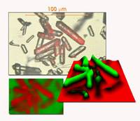
Raman Mapping Horiba. Generating confocal images becomes faster easier and more flexible from the deep UV to the IR. In Biology and Life Sciences it can be used to image tissues whole cells or their components without the need for dyes and stains or to locate Raman and SERS tags.
In Material Sciences it can investigate how structure stress and strain vary across a sample. Ruko Jalur Sutera Jl. LabRAM Soleil New Confocal Raman Microscope from HORIBA Scientific Your ideal lab companion.
The result of 50 years of expertise in Raman spectroscopy LabRAM Soleil offers unprecedented capabilities for Raman multimodal confocal imaging in a compact footprint.
A complete spectrum is acquired at each and every pixel of the image and then interrogated to generate false color images based on material composition and structure. Thus whether the application is for routine screening fluorescence and PL mapping or detailed high resolution Raman research the spectrometer to match is available. In Material Sciences it can investigate how structure stress and strain vary across a sample. A complete spectrum is acquired at each and every pixel of the image and then interrogated to generate false colour images based on material composition and structure.
